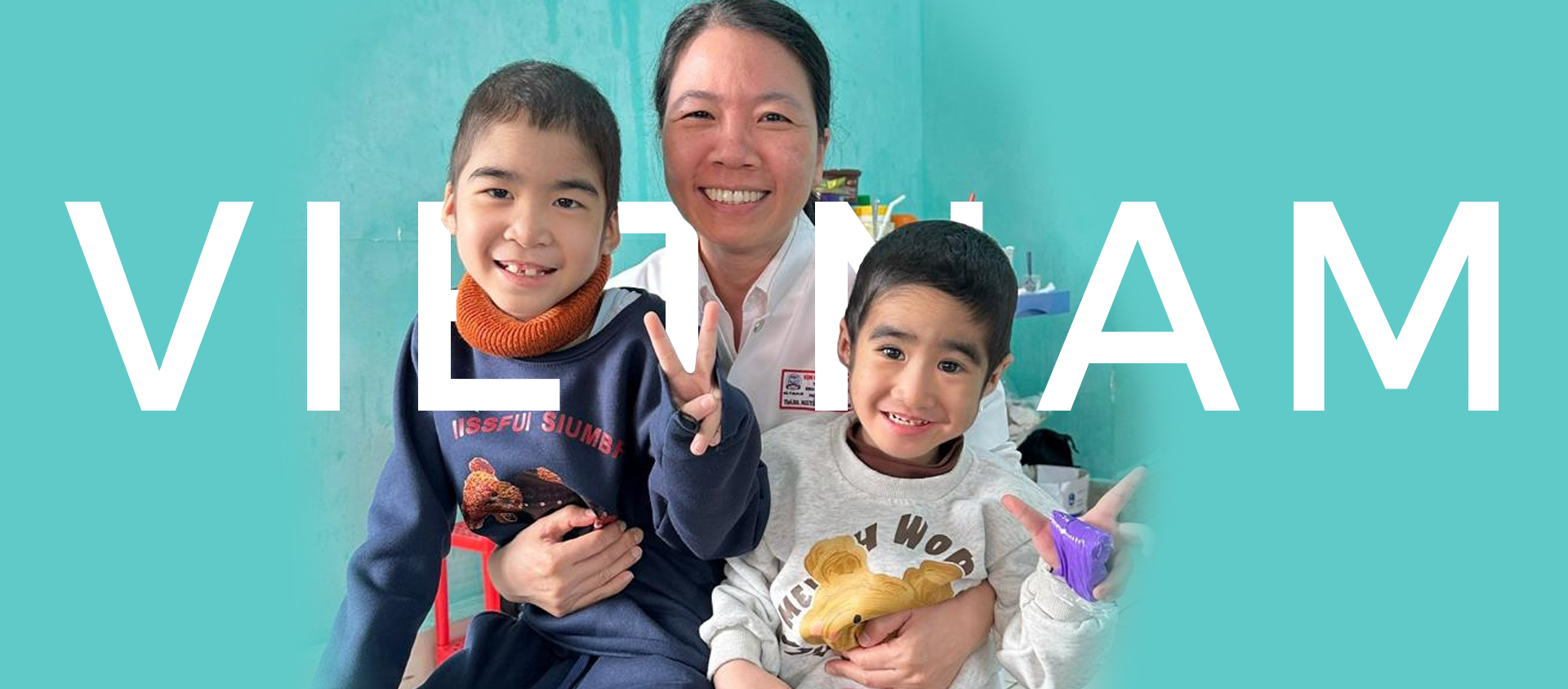
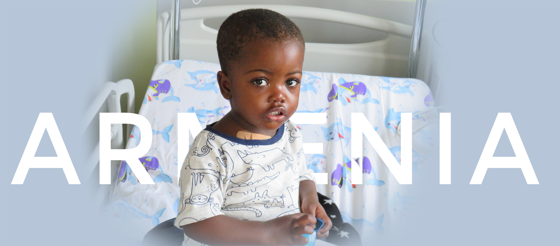
Navigation

Navigation

Rhabdomyosarcoma (RMS)


Accounts for 50% of soft tissue sarcomas in the pediatric age group. The estimated average annual incident rate is 4.3 cases per million children, 2/3 are less than 6 years old and the remaining third are adolescents. It is slightly more frequent in males1.

Pathology

Small round blue cell tumor with skeletal myogenic features on morphology, immunohistochemistry (muscle-specific actin and myosin, desmin, myoglobin, Z-band protein, and MyoD), and molecular genetics.

Alveolar (ARMS): more frequently adolescents, extremities, in ΒΌ of cases T(2;13)(q35;q14) (PAX3-FKHR) is present.

Embryonal (ERMS): generally younger than 6 years and in central locations (e.g orbit). In over 50% of cases there if loss of heterozygosity at 11p15 (where the IGF2 gene is located). Botroyd is a favourable subtype of embryonal.

A standardized protocol for handling tissue specimens has been developed 2.

Minimal disease evaluation

Even though immunocytology for MyoD (muscle differentiation factor widely expressed in RMS), Desmin, or PCR for ARMS :T(2;13)(q35;q14) (PAX3-FKHR) have been used, there is no methodology which has been validated and shown to be clinically useful.

Pattern of spread and staging studies

As a consequence, approximately 25% of patients may have evidence of metastatic disease: lung 50%, bone marrow 30%, lymphnodes 20% bone 10%. Visceral mestastasis such as liver or brain are generally seen in preterminal patients. Some patients with metastatic disease such as ARMS histology PAX7-FKHR variant translocation or ERMS and <10 years may do well with EFS in the range of 50% 3.

Initial work up

Routine evaluations: blood chemistries (electrolites, Ca, P, Mg, BUN, serum creatinine, uric acid, total bilirubin, transaminase, PT, PTT, fibrinogen, complete blood count), urine analysis, chest xRay and abdominal ultrasound.

Primary site imaging (MRI, CT or sonography depending on available techniques), MRI, Lung CT, Bone marrow aspirate and biopsy in two sites, skeletal x-ray survey.

Isolated BM metastasis are rare (<2%), the value of trephine biopsies in otherwise M0 patients is questionable and a bone marrow aspirate is probably adequate.

A bone scan may not be an absolute necessity; in RMS patients bone scans may not always be more sensitive than plain x-ray 4. Very few patients are picked up by bone scans that have negative routine skeletal survey or BM aspirates.

Brain imaging is not necessary for tumors arising outside the head and neck. While parameningeal tumors should be evaluated with an exploratory LP in addition to CT or MRI.

A baseline echocardiogram should be obtained for patients undergoing therapy with anthracyclins.

Risk factors and prognostic stratification

Genetic aberrations do not currently impact on the risk-group assignment.

Histology: ARMS unfavourable, in the IRS III study, however, more intensive therapy reduced the value of histology.

Site: Low risk: orbit, eyelids, head and neck, non-paramengeal, genitourinary tract (except bladder or prostate). High-risk: extremity, bladder, prostate, trunk, retroperitoneal, parameningeal.

Margins

Limphonodes

Metastasis

Treatment response

RMS risk group assignment algorythm

see also attached tables

Treatment

Surgery should be performed, keeping in mind that there is little evidence that aggressive approaches greatly impairing function or cosmesis are justified. Second look surgery after chemotherapy to convert a partial response into a complete response or to verify complete radiological remission and avoid radiotherapy can be misleading due to a high rate of false positivity and may not impact on the final outcome 5.

Lymphnode spread is quite rare with head and neck ERMS lesions (<5%) as opposed to extremity and paratesticular tumors (in boys of around 10 years) where it can reach 50%. In this latter circumstance ipsilateral lymph node dissection is warranted even if nodes are clinically/radiologically negative or doubtful. An enlarged node should always be resected.

Pulmonary metastectomy in patients with RMS is not justified both at diagnisis and at recurrence 6.

Radiotherapy is generally administered in doses of 40 Gy in 180-200 cGy daily fractions to sites of microscopic residual disease or lymphnode involvement. Doses higher than 50 Gy may be used for gross residual disease. Treatment fields should encompass the pre-treatment tumor volume with a 2 cm clear margin. Even in lesions <5 cm with negative margins RT should probably be administered to ARMS histologies.

The value of whole lung radiation with cumulative doses of 14,4 Gy for lung metastases is uncertain as well as the role of intraoperative RT (IORT).

Radiotherapy can be postponed up to 12 weeks from diagnosis while chemotherapy is administered. For parameningeal tumors, however, RT should be started within 2 weeks from diagnosis 7. There is no value in whole brain RT for meningeal spread or intracranial extension 8.

Chemotherapy

With local control only, the great majority of patients eventually metastasise and die.

Chemotherapy can generally begin after one week from surgery and should not be postponed beyond 8 weeks. The most active agents are dactinomycin, cyclophosphamide, vincristine and doxorubicin. Other active agents include ifosfamide, cisplatin, etoposide, melphalan and dacarbazine. Topotecan, irinotecan and vinerolbine are newer drugs with promising activities.

Stage 1: completely resected, treatment may be limited to a 2 drug regimen: VA, i.e. vincrisitne 1.5 mg/m2 weekly, with a maximum dose of 2 mg, and dactinomycin 1.5 mg/m2, with a maximum dose of 2 mg every 3 weeks, followed by 3 weeks rest, this is given for 3 times for a total of 6 doses of dactimomycin. Radiotherapy maybe avoided.

Stage 2: the gold standard combination is VAC, i.e. vincristine, dactinomycin and cyclophosphamide (1,200 mg/m2 in 1 day at 4 weeks intervals). The minimum duration of therapy is not known but is probably not less than 9 cycles, i.e approximately 27 weeks. Radiotherapy maybe avoided if complete resection is well documented with the possible exception of ARMS histologies.

Stage 3 and 4 patients should be treated with intensive standard VAdrC alternating with VAC for a total 12-14 cycles, the optimal duration of therapy is not known but there is no evidence that longer treatments are of any benefit. Also, the addition of etoposide may not be justified9. Most patients older than 3 years with microscopic or gross residual tumor should receive local RT, if no local radiotherapy is administered, intensified chemotherapy should be considered. For stage 4 patients primary site RT should be administered only if metastatic disease is in CR or VGPR at week 9 evaluation.

Ifosfamide does not seem to offer any advantage over cyclophosphamide.

The role of autologous stem cell transplantation is unclear, if an autologous rescue is available it might be reasonable to use high-dose melphalan (100-120 mg/mq) in combination with standard dose vincristine for 1 or 2 cycles 4 weeks apart, with the aim of decreasing toxicity and therapy duration rather that delivering supramaximal chemotherapy. In fact, in the contexts in which transfusions may hardly be available, or if treatment abandonment is likely, melphalan with autologous stem cells may be associated with minimal transfusion requirements, very short neutropenias, and therapy may be completed in 12 weeks.

Notes

Avoid the use of cyclophosphamide in patients who received bladder RT

Dactinomyicin should not be administered concomitantly with RT.

Special cases

A minority of cases may have special features that will justify a case by a case decision making process:

Children younger than 1 year (<5%), may have a worse prognosis independently of other risk factors.

In children younger than 3 years RT should be avoided even in the presence of residual disease in favour of more aggressive surgery and/or chemotherapy.

Even if metastatic, patients <10 years with ERMS and those with variant translocation PAX7-FKHR ARMS may do well (EFS 50%) and should be treated aggressively.

Treatment response assessment and follow up

All measurable lesions should be evaluated clinically after each cycle, while radiological diseases status assessment should be performed prior to the fourth chemotherapy cycle (approximately week 9), after the completion of radiotherapy (approximately week 17) and at the end of therapy (approximately week 27).

Response definition: CR (complete response, no measurable disease), VGPR (very good partial response, >90% reduction of maximal tumor diameter), PR (partial response, >50% reduction), NR (no response, <50% reduction), PD (progressive disease, increase in size or metastatic spread). A recurrence should be called such only after achieving a previous CR or VGPR.

Relapses after 10 years are very rare, more than 90% will occur within the first 3 years from diagnosis.

The curability of a relapse depends on the initial stage, prior therapy intensity, time to relapse, and whether the recurrence is local or distant. Thus, a patient's condition, when the patient received low-intensity therapy and/or no local RT, should be followed particularly carefully for the first 3 years from diagnosis focussing on primary site evaluation, with routine physical examinations and blood test evaluations as well as local US and a chest ray every 4 months. Patients relapsing after intensive chemotherapy and appropriate local control measures or at distant sites have a very little chance of being cured.

References

  1. Pizzo AP, Poplack DG. Principles and practice of pediatric oncology. Philadelphia: J.B.Limpicott Company; 2006.
  2. Qualman SJ, Bowen J, Parham DM, Branton PA, Meyer WH. Protocol for the examination of specimens from patients (children and young adults) with rhabdomyosarcoma. Arch Pathol Lab Med. 2003;127:1290-1297.
  3. Sorensen PH, Lynch JC, Qualman SJ, et al. PAX3-FKHR and PAX7-FKHR gene fusions are prognostic indicators in alveolar rhabdomyosarcoma: a report from the children's oncology group. J Clin Oncol. 2002;20:2672-2679.
  4. Quddus FF, Espinola D, Kramer SS, Leventhal BG. Comparison between x-ray and bone scan detection of bone metastases in patients with rhabdomyosarcoma. Med Pediatr Oncol. 1983;11:125-129.
  5. Godzinski J, Flamant F, Rey A, Praquin MT, Martelli H. Value of postchemotherapy bioptical verification of complete clinical remission in previously incompletely resected (stage I and II pT3) malignant mesenchymal tumors in children: International Society of Pediatric Oncology 1984 Malignant Mesenchymal Tumors Study. Med Pediatr Oncol. 1994;22:22-26.
  6. Temeck BK, Wexler LH, Steinberg SM, McClure LL, Horowitz M, Pass HI. Metastasectomy for sarcomatous pediatric histologies: results and prognostic factors. Ann Thorac Surg. 1995;59:1385-1389.
  7. Michalski JM, Meza J, Breneman JC, et al. Influence of radiation therapy parameters on outcome in children treated with radiation therapy for localized parameningeal rhabdomyosarcoma in Intergroup Rhabdomyosarcoma Study Group trials II through IV. Int J Radiat Oncol Biol Phys. 2004;59:1027-1038.
  8. Crist W, Gehan EA, Ragab AH, et al. The third Intergroup Rhabdomyosarcoma Study. J Clin Oncol. 1995;13:610-630.
  9. Dantonello TM, Int-Veen C, Harms D, et al. Outcome of children and adolescente with localized rhabdomyosarcoma treated according to the cooperative soft tissue sarcoma study CWS-91 [abstract]. Pediatr Blood Cancer. 2006;47:O.056.
  10. Meza JL, Anderson J, Pappo AS, Meyer WH. Analysis of prognostic factors in patients with nonmetastatic rhabdomyosarcoma treated on intergroup rhabdomyosarcoma studies III and IV: the Children's Oncology Group. J Clin Oncol. 2006;%20;24:3844-3851.
AttachmentSize
Rhabdomyosarcoma_tables and figuresRM.pdf88.39 KB


How to help us


Birthday Solidarity
Become a Volunteer
Donation
5X1000
C.F. 05712190486


Newsletter

Newsletter

User login